

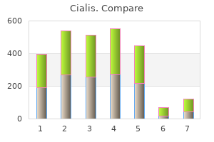
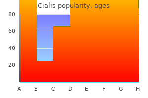
Cialis
"Buy cialis 5mg on-line, impotence remedies".
By: U. Kalan, M.B. B.CH., M.B.B.Ch., Ph.D.
Professor, University of Illinois College of Medicine
These questions relate to features that have been shown to put depressed individuals at risk of suicide erectile dysfunction drugs india buy cialis 10mg amex. If erectile dysfunction treatment youtube discount 20 mg cialis with mastercard, from their answers erectile dysfunction drug coupons cialis 5mg with amex, they are judged to carry an imminent risk of suicide impotence merriam webster generic 10 mg cialis otc, they should be directed to a psychiatrist and generally admitted to a hospital. In some depressions, hypochondriacal preoccupation with bowel and digestive functions accounts for repeated visits to the medical clinic. In one study, 21 of 120 such patients were subsequently diagnosed as being depressed. Early awakening is typical, and the morning hours are then the worst period of the day. Other patients have difficulty falling asleep, especially if there is an associated anxiety state. A complaint in the male of loss of libido and impotence is another monosymptomatic presentation; only with probing inquiry about other disturbances common to depression will the diagnosis become evident. The manic state is, in most ways, the opposite of the depressed state, being characterized by a flight of ideas, motor and speech hyperactivity, and an increased appetite and sex urge. The manic individual appears to possess great drive and confidence yet lacks the ability to carry out plans. Judgment may be so impaired that reckless investments are made; fortunes are spent in gambling or on extravagant shopping sprees. Euphoria and expansiveness sometimes bubble over into delusions of power and grandeur, which, in turn, may make the patient offensively aggressive. The threshold for paranoid thinking is low, which makes the patient sensitive and suspicious. In its most advanced form, a condition described as "delirious mania," the patient becomes totally incoherent and altogether disorganized in behavior. At this stage visual and auditory hallucinations and paranoid delusions may be rampant; furthermore, as the term delirium implies, the patient may be disoriented and agitated, with a clouded sensorium. As mentioned earlier, one observes patients with repeated attacks of mania without depression at any time, but this is not common. Hypomania represents a milder degree of the disorder, but this term is also used loosely to depict behavior in a normally functioning individual who is unusually energetic and active. In this latter sense hypomania is a personality trait found in many talented and productive persons and need not arouse concern unless it is excessive and out of character for the individual. As commented in the review by Belmaker, a number of highly creative individuals have had bipolar disorders, but full-blown mania is uniformly destructive of careers and personal relationships. He cites Shou in pointing out that such individuals are actually more creative when treated with appropriate medications. First attacks of either depression or mania last an average of 6 months if untreated, although the duration varies greatly. Although most attacks of manic-depressive disease subside in a matter of months, a significant number, unipolar patients more than bipolar ones, remain chronically ill for long periods. According to Winokur and colleagues, 14 percent of their bipolar patients had not recovered after 2 years and 5 percent after 5 years. Comparative figures for primary unipolar patients were 19 and 12 percent, respectively. Variables that are predictive of an unfavorable outcome are high degrees of neuroticism, strongly positive family history of a similar psychiatric illness, and presence of depressionprovoking circumstances (Hirschfeld et al). In this diagnostic scheme, each of the four diagnostic symptoms should have been present for at least 2 weeks. As mentioned earlier, depressed patients who are referred to the neurologist tend to complain inordinately of physical and cognitive symptoms and to minimize or deny the purely affective ones. Complaints of fatigue, weakness, malaise, or widespread aches and pains, for example, suggest a variety of medical diseases, such as Addison disease, hypothyroidism, chronic infection, polymyositis, or early rheumatoid arthritis. Often the fatigue state is misinterpreted as muscular weakness, and this directs a medical search for neuromuscular disease.
A purely myopathic form of hypertonia is difficult to substantiate (the exception being hypocalcemic tetany) erectile dysfunction drugs viagra order cialis 20 mg without a prescription. Various types of disease may lead to fibrous contracture and arthrogryposis erectile dysfunction medication risks generic 5mg cialis, as stated earlier erectile dysfunction kya hai cialis 20mg amex. For the most part erectile dysfunction blue pill buy genuine cialis line, the muscle stiffness syndromes are due to continuous overactivity of motor units, the extreme forms being the stiff-man and the Isaacs syndromes and several related conditions (Chap. Changes in Muscle Volume Diminution or increase in muscle bulk stands as another feature of disease that can be observed in all except the most obese patients. There are, of course, innate differences in muscle development, a greater salience of muscle in the male than in the female, and differences due to use and disuse. In general, the relative size of muscles is a genetic trait; in some families, muscles, like the skeleton, are large and in others, small. Greatly increased size and strength of muscles (hypertrophia musculorum vera) is an inherited trait caused by a mutation in the myostatin gene. It also is observed in cultists devoted to bodybuilding (although, as a rule, they are exercising muscles that are large from birth); in congenital myotonia (circus freaks with phenomenal muscular development often have this condition); in instances of a rare pathologic cramp syndrome; in the Bruck-DeLange syndrome of congenital hypertrophy of muscle, athetosis, and mental retardation; and in some patients destined to develop muscular dystrophy. In some of the latter patients, the muscle enlargement is in reality a pseudohypertrophy, in which the increase in size is accompanied by weakness. Here large and small fibers are mixed with fat cells, which have replaced many of the degenerated muscle fibers; other muscles in the same patient are atrophied. Rarely, we have seen it in amyloidosis, sarcoidosis, eosinophilic myositis, and certain of the congenital myopathies. Hypothyroidism is often accompanied by an increase in volume of certain muscles, at times pronounced enough to simulate the hypertrophia musculorum vera. Enlargement of muscles in only one limb may be due to an arteriovenous malformation, neurofibroma, or the Klippel-Trenaunay-Weber syndrome involvґ ing the nerve root. A single muscle may become hypertrophied after partial injury to its nerve root (see Mielke et al); this is seen most often in relation to disc compression or surgical injury of the S1 nerve root. Lesions of the peripheral nerve or anterior horn cells, if complete, lead to a loss of bulk of up to 85 percent of the original volume within 3 to 4 months, although some shrinkage of muscle becomes evident before that time. The most severe degrees of atrophy are observed in the chronic polyneuropathies, motor system diseases, and muscular dystrophies. Of interest is the fact that a number of myopathic diseases result in severe weakness with little or no Muscle Tone All normal muscles display a slight resistance to stretch even when fully relaxed. When stretched to their limit and released, they recoil, mainly because of the elasticity of the fibers and their connective tissue sheaths. In addition, the trunk and proximal limb musculature is intermittently or constantly activated in the maintenance of attitudes and postures. A reduced innervation of muscle, atrophy, or loss of contracting fibers are causes of "hypotonia. Tone can be assessed by lifting the infant in a prone position; if the child is hypotonic, the head and legs cannot be supported against gravity and, therefore, dangle. Andre-Thomas and colleagues introduced ґ the terms passivite, to refer to the amplitude of the flapping moveґ ment of the hand or foot when the limb is shaken, and extensibilite, ґ to denote the reduced resistance of a relaxed muscle to slow passive stretch. Diminution in tone is observed in infants suffering from Werdnig-Hoffman disease or the congenital myopathies as well as in those with a benign type of hypotonia; diminished tone may also be seen in sickly infants with a variety of systemic diseases. Twitches, Spasms, and Cramps Fascicular twitches of muscle at rest (fasciculations), if pronounced and combined with muscular weakness and atrophy, usually signify motor neuron disease (amyotrophic lateral sclerosis, progressive muscular atrophy, or progressive bulbar palsy); but they may be seen in other diseases that involve the gray matter of the spinal cord. Widespread fasciculations may occur acutely with severe dehydration and electrolyte imbalance after an overdose of neostigmine or with organophosphate poisoning. Slow and persistent fasciculations, spreading in a wave-like pattern along the entire length of a muscle and associated with slight reduction in the speed of contraction and relaxation, are part of the syndrome of continuous muscular activity (page 1278). Fasciculations that occur with muscular contraction, in contrast to those at rest, indicate a state of heightened irritability of muscle; this may occur for no discernible reason or as a sequela of denervation that leaves muscle with some paralyzed motor units, so that during contraction small and increasingly larger units are not enlisted smoothly. Fasciculations of the facial muscles are typical of the Kennedy type of bulbospinal atrophy. Benign fasciculations, a common finding in otherwise normal individuals, can be identified by the lack of muscular weakness and atrophy and by the smallness of the muscle fascicle involved and repetitive appearance in only one or two regions. The recurrent twitches of the eyelid or muscles of the thumb experienced by most normal persons are often referred to as "live flesh" or myokymia but are benign fasciculations of this type.
Purchase cialis canada. Homeopathic medicine for Premature Ejaculation! Erectile dysfunction !.
Flat palpation is used to press fingertip portions of the orbicularis oculi against the underlying bony orbit which antihypertensive causes erectile dysfunction generic cialis 2.5 mg free shipping. These two muscles are important not only for facial expression but also in ocular reflexes erectile dysfunction and diabetes leaflet order cialis 2.5 mg with visa. When it contracts erectile dysfunction causes agent orange order cheap cialis on line, it enlarges the nostrils and elevates the nasal wing impotence medication buy genuine cialis online, producing transverse folds in the skin on each side of the nose and a look of displeasure and discontent, especially noted when sniffing an unpleasant odor. Since this is an action people often perform when experiencing a headache or eyestrain, its association with those patterns of dysfunction may be implied. Flat palpation and light friction may be used along the sides of the nose and spreading slightly laterally onto the cheeks to treat the remaining nasal muscles. The two index fingers, very lightly placed, may provide precise myofascial release but the practitioner is reminded that the facial tissues are very delicate and anything other than exceptionally light pressure is contraindicated. Trigger point locations and patterns of referral in this region have not yet been established but we suggest that these muscles be assessed when nose, lips and eye problems are encountered or facial pain or sensations are experienced near or into these tissues. Wrinkled skin may suggest underlying muscular tensions, possibly involving chronic overuse. Gentle static pressure or an extremely gentle transverse movement may help assess the underlying muscle. However, frictional movements, gliding techniques or skin rolling, which is usually effective in locating trigger points, may also be too aggressive for this delicate tissue. The corrugator supercilii is easily picked up near the mid-line between the brows and compressed between the thumb and side of the index finger. This compression and rolling technique is applied at thumb-width intervals the width of the brow and may also include fibers of the procerus, frontalis and orbicularis oculi as well as corrugator supercilii. It reduces glare from excess light and produces transverse wrinkles at the bridge of the nose. Expressions associated with procerus include menacing looks, frowns and deep concentration. Nasalis consists of a transverse (compressor naris) portion which attaches the maxilla to the bridge of the nose and an alar (dilator naris) portion which attaches the maxilla to the skin on the nasal wing. The transverse portion compresses the nasal aperture while the alar portion widens it, reducing the size of the nostril and producing a look of desiring, demanding and sensuousness. Depressor septi attaches the mobile portion of the nasal septum to the maxilla above the central incisor tooth. Elevators, retractors and evertors of the upper lip: levator labii superioris alaeque nasi, levator labii superioris, zygomaticus major and minor, levator anguli oris and risorius Depressors, retractors and evertors of the lower lip: depressor labii inferioris, depressor anguli oris and mentalis Compound sphincter: orbicularis oris, incisivus superior and inferior Buccinator the muscles of the buccolabial region function in eating, drinking and speech as well as emotional expression. A multitude of expressions, including reserve, laughing, crying, satisfaction, pleasure, self-confidence, sadness, perseverance, seriousness, doubt, indecision, disdain, irony and a variety of other feelings, are displayed in the lower face by the action and combined actions of these muscles. A number of muscles of the buccolabial region converge into the modiolus just lateral to the buccal angle of the mouth. The modiolus can be palpated in an intraoral examination and is usually felt as a dense, mobile fibromuscular mass that may or may not be tender. This fan-shaped radiation of muscular fibers allows the three-dimensional mobility of the modiolus to integrate facial activities of the lips and oral fissure, cheeks and jaws, such as chewing, drinking, sucking, swallowing and modulations of various vocal tones. The practitioner should wear protective gloves see precautions for intraoral examination on p. The index finger of the gloved treatment hand is placed inside the mouth and the thumb is placed on the outside (facial) surface. The tissue is compressed between the two digits as the internal finger is slid against the external thumb while manipulating the tissue held between them. The treating digits progress at thumb-width intervals around the mouth until all the tissues have been examined. Tender spots or trigger points may be treated with static pressure; alternatively, spray and stretch techniques, as described by Simons et al (1999), may be used with precautions as noted in their text. The buccolabial muscles may also be treated from an external perspective by pressing them against the underlying maxilla, mandible or teeth and flat palpation can be used to assess and treat them.
These neurons project to pyramidal neurons in layer 5 treatment for erectile dysfunction before viagra cheap cialis 5 mg line, which in turn activate pyramidal neurons in layer 6 erectile dysfunction treatment in rawalpindi buy cialis pills in toronto. The excitatory interactions within a column are controlled by different types of local interneurons (not shown) erectile dysfunction onset buy 2.5 mg cialis otc, which also mediate lateral inhibition of surrounding columns cialis causes erectile dysfunction discount cialis. The apical dendrites of pyramidal cells reach layer 1, where they receive input from other cortical areas, thalamic intralaminar nuclei, and cholinergic and monoaminergic systems of the brainstem, hypothalamus, and basal forebrain. Cortical pyramidal cells give rise to extrinsic connections: layer 2 cells to intrahemispheric corticocortical association fibers, layer 3 cells to interhemispheric commissural fibers, layer 5 cells to corticostriate, corticorubral, corticopontine, corticobulbar, and corticospinal fibers, and layer 6 cells to corticothalamic fibers. Most studies using brain anatomy use the Brodmann classification scheme, and, depending on the area of inquiry, other regions are often mentioned. Finally, although poorly understood, there is an important laterality effect such that the left hemisphere in essentially all right-handed persons and in roughly 75% of left-handed persons supports language, whereas the right hemisphere in such cases appears to be dominant for spatial skills. Some left-handed persons have mixed language dominance, and a small subset are completely right hemisphere language dominant. Neuroscience and Neuroanatomy Primary sensory cortex Unimodal sensory association cortex Heteromodal association cortex Paralimbic cortex Limbic cortex Primary motor cortex Premotor cortex Hypothalamus Extrapersonal space Internal milieu Figure 21. Sensory information is processed serially from primary sensory, to unimodal sensory, to heteromodal sensory association areas in the posterior parietal and lateral temporal cortices. These areas project to both heteromodal association areas of the frontal lobe (prefrontal cortex) and paralimbic areas, which provide input to the hippocampus and amygdala. The prefrontal cortex projects to premotor areas (unimodal motor association areas), which activate primary motor cortex. Note the feedback connections between the paralimbic, heteromodal, and unimodal areas. The cortex then projects back to wide regions of the central nervous system including the cortex, thalamus, basal nuclei, cerebellum, brainstem, and cord. The types of fibers connecting areas of the central nervous system are designated based on regions they connect. Multiscale Organization the human brain is a complex information processing system whose proper functioning depends on the organization of its processing units. The scale of these processing units spans multiple levels from the genetic to systems level (Figure 22. A complete understanding of any 1 of these levels necessitates understanding each level because they are interdependent. For example, the genetic expression of a signal neuron determines the protein composition of that neuron, which, in turn, influences its firing pattern within a microcircuit. Microcircuits form local circuits that interact to form larger regional circuits and eventually influence systems-leveldistributed neural networks that are more closely associated with behavioral interactions with the environment. Thus, the firing pattern of neurons within circuits changes, which leads to a change in the genetic expression within individual neurons in order to optimally respond to this new firing pattern. The complex interaction between these various levels is typically summarized as an interaction between genes and environment. At any point in time, it is impossible to know every piece of information necessary to fully characterize the complexity within these systems. However, general principles of this multiscale organization are emerging via interdisciplinary investigations of complex adaptive systems. These principles can be more easily characterized and used to understand the brain in health and disease. A discussion of these complex network principles is beyond the scope of this chapter, but the structural, functional, and behavioral aspects of this multiscale system are reviewed in more detail. Therefore, the basic bipartite functional unit (ie, 2 neurons) is supported by a basic tripartite structural unit (ie, neuron, astrocyte, and a vascular endothelial cell). Neurovascular coupling related to this bipartite functional and tripartite structural arrangement allows for the identification of regions of the brain that are functionally connected at the systems level. The laminar organization of the cortex is the basic structural substrate of cortical functional units at the microcircuit level. The cerebral cortex is composed of 6 possible cortical layers, with regional variability in the presence of these layers and their cellular composition, commonly referred to as the cytoarchitectonics of the region (see Chapter 21, "Cortex Topography and Organization"). Anterior and Posterior Limbic Circuits: Emotion and Memory the structures involved in the anterior and posterior limbic systems are evolutionarily primitive, which speaks to their critical role in fundamental aspects of behavior, with higher neocortical functions arising with the need to modulate these primitive circuits. The brain evolved only once in evolutionary history, and all subsequent organisms with nervous systems share a common ancestor. Bilaterally symmetric mobile muticellular organisms driven to interact with the extrapersonal environment first developed a primitive olfactory sensory system in order to efficiently guide movements to sources of sustenance.