

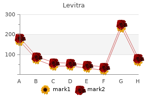
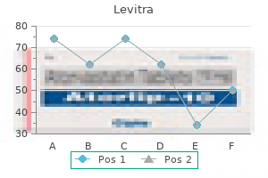
Levitra
"Buy levitra visa, erectile dysfunction drugs nhs".
By: T. Kafa, M.S., Ph.D.
Professor, University of Washington School of Medicine
Equipment Iodine-131 therapy is sometimes carried out impotence blood circulation order levitra 20 mg, especially in patients suspected to have metastatic cancer erectile dysfunction diabetes buy generic levitra 20 mg on-line, after demonstration of iodine-avid thyroid tissue (normal or malignant) by a gamma camera or whole body counter erectile dysfunction melanoma discount generic levitra canada. Most centres carry out gamma camera imaging using a high energy erectile dysfunction treatment michigan order levitra uk, general purpose collimator. Most centres also carry out imaging with comparable imaging methods, to demonstrate targeting of therapeutic 131I to thyroid tissue. No special equipment is required for outpatient therapy, apart from adequate shielding of the 131I and appropriate monitoring of patients to ensure adherence to radiation safety criteria for outpatient therapy. High doses of 131I should be administered within areas that meet radiation protection requirements. Radiopharmaceuticals Iodine-131, in the form of sodium iodide, is administered orally. Action prior to 131I therapy Patients at intermediate or high risk of thyroid cancer usually receive 131I therapy after definitive thyroid surgery (usually total or radical thyroidectomy, with recurrent laryngeal nerve and parathyroid preservation). Skin sterilization for thyroid surgery must not use an iodine containing compound. Patients must not receive thyroid hormone replacement for at least four weeks prior to 131I therapy. Patients who tolerate hormone withdrawal poorly may receive tri-iodothyronine (T3) until two weeks prior to therapy. No intravenous contrast should be administered for at least two months prior to planned evaluation and therapy. Patients should be encouraged to reduce the iodine content in their diet to optimize uptake of 131I by thyroid tissue. Serum thyroglobulin estimations are usually carried out immediately prior to administration of 131I tracer. A tracer study may be carried out prior to administration of 131I therapy, to ensure 131I uptake in thyroid tissue and/or in metastatically diseased tissue. Whole body imaging at 72 hours should also be carried out, especially when the results of neck imaging are negative. A form signed by the patient giving their informed consent for therapy is required. Therapy Ablative therapy is defined as that given immediately following definitive surgery. When the mass of thyroid remnant can be estimated, for example using ultrasound, a dose of 131I calculated to deliver 3050 Gy to the thyroid remnant may also be used. Ablative therapy should be given to all patients with iodine-avid thyroid/malignant tissue in the neck or elsewhere, or in those patients who, immediately after surgery, have no evidence of iodine-avid thyroid tissue 72 hours after oral administration of 131I tracer but who have elevated serum thyroglobulin levels. Patients should be evaluated not earlier than six months after ablative 131I therapy for evidence of residual or recurrent disease. This evaluation is carried out not less than four weeks after cessation of thyroid hormone replacement or, if the patient cannot tolerate hormone withdrawal, by the following regimen: - Stop levothyroxine and substitute with a comparable dose of T3 for two weeks. Anterior and posterior whole body imaging should be carried out at least 72 hours after administration of the tracer, using high energy collimation. An alternative to whole body imaging is static anterior and posterior imaging of the relevant areas (head, neck, chest, abdomen, pelvis and lower extremities), taken for at least 10 min each. If there is evidence of iodine-avid disease from scintigraphy and/or if the serum thyroglobulin level is elevated, the patient should be treated with 131I. The maximum safe dose of 131I has been found to be that which delivers no more than 2 Gy to the blood. Post-therapy follow-up Hormone replacement may be resumed two days after treatment. In most centres, anterior and posterior images of the body are obtained a week to 10 days after 131I therapy to ensure targeting. This can be done most reliably when the patient is no longer on T4 or T3 treatment. When patients are treated at the maximum safe dose, haematological evaluation should be carried out between four and six weeks after therapy, to ensure lack of haematopoietic toxicity. Patients are usually not re-treated earlier than six months after therapy, unless there is evidence of rapidly progressive disease as evidenced by a progressive rise in serum thyroglobulin and/or radiographic evidence of progressive disease. Two successive negative whole body studies, with concurrent non-measurable serum thyroglobulin levels, separated by intervals of at least six months, indicate successful therapy. The patient may then be managed by serum thyroglobulin estimations twice yearly for five years and then annually for at least another five years.
The spinalis group includes the spinalis capitis erectile dysfunction treatment exercise purchase levitra 20mg without a prescription, the spinalis cervicis erectile dysfunction etiology cheap 20 mg levitra amex, and the spinalis thoracis impotence of organic nature purchase cheap levitra. The transversospinales include the semispinalis capitis erectile dysfunction pumps side effects order 10 mg levitra otc, semispinalis cervicis, semispinalis thoracis, multifidus, and rotatores. Finally, the scalenes include the anterior scalene, middle scalene, and posterior scalene. These muscles include the rectus abdominis, which extends through the entire length of the trunk, the external oblique, the internal oblique, and the transversus abdominus. The muscles of the thorax play a large role in breathing, especially the dome-shaped diaphragm. When it contracts and flattens, the volume inside the pleural cavities increases, which decreases the pressure within them. The external and internal intercostal muscles span the space between the ribs and help change the shape of the rib cage and the volume-pressure ratio inside the pleural cavities during inspiration and expiration. The perineum muscles play roles in urination in both sexes, ejaculation in men, and vaginal contraction in women. The pelvic floor muscles support the pelvic organs, resist intra-abdominal pressure, and work as sphincters for the urethra, rectum, and vagina. The muscles that position and stabilize the pectoral girdle are located on the thorax. The anterior thoracic muscles are the subclavius, pectoralis minor, and the serratus anterior. The posterior thoracic muscles are the trapezius, levator scapulae, rhomboid major, and rhomboid minor. The ones that originate on the axial skeleton are the pectoralis major and the latissimus dorsi. The deltoid, subscapularis, supraspinatus, infraspinatus, teres major, teres minor, and coracobrachialis originate on the scapula. The extrinsic muscles of the hands originate along the forearm and insert into the hand in order to facilitate crude movements of the wrists, hands, and fingers. These muscles are the flexor carpi radialis, palmaris longus, flexor carpi ulnaris, and the flexor digitorum superficialis. The extensor carpi radialis longus, extensor carpi radialis brevis, extensor digitorum, extensor digiti minimi, and extensor carpi ulnaris are the muscles found in the superficial posterior compartment. Finally, the intrinsic muscles of the hands allow our fingers to make precise movements, such as typing and writing. The thenar muscles, which are located on the lateral part of the palm, are the abductor pollicis brevis, opponens pollicis, flexor pollicis brevis, and adductor pollicis. The hypothenar muscles, which are located on the medial part of the palm, are the abductor digiti minimi, flexor digiti minimi brevis, and opponens digiti minimi. The intermediate muscles, located in the middle of the palm, are the lumbricals, palmar interossei, and dorsal interossei. The large and strong gluteus maximus, gluteus medius, and gluteus minimus extend and abduct the femur. Along with the gluteus maximus, the tensor fascia lata muscle forms the iliotibial tract. The lateral rotators of the femur at the hip are the piriformis, obturator internus, obturator externus, superior gemellus, inferior gemellus, and quadratus femoris. On the medial part of the thigh, the adductor longus, adductor brevis, and adductor magnus adduct the thigh and medially rotate it. The thigh muscles that move the femur, tibia, and fibula are divided into medial, anterior, and posterior compartments. The anterior compartment comprises the quadriceps femoris, quadriceps tendon, patellar ligament, and the sartorius. The quadriceps femoris is made of four muscles: the rectus femoris, the vastus lateralis, the vastus medius, and the vastus intermedius, which together extend the knee.
Both diseases are autosomal dominant erectile dysfunction remedies order levitra mastercard, but spontaneous mutations account for approximately half of cases erectile dysfunction 43 purchase 10 mg levitra overnight delivery. The incidence of neurofibromatosis type 1 is 1 in 2500; the mutated gene product is neurofibromin erectile dysfunction drugs injection generic levitra 10mg with mastercard, a protein involved in tumor suppression erectile dysfunction treatment urologist levitra 20mg on-line. Neurofibromatosis type 2 has a reported incidence of 1 in 33,000; the involved gene product is merlin, which mediates cytoskeleton and extracellular movement. Neurofibromatosis type 1 should be suspected in any infant with multiple cafй-au-lait spots, congenital glaucoma, a plexiform neurofibroma, or pseudoarthrosis. Without a positive family history, however, it can be difficult to diagnose neurofibromatosis in the first months of life. The diagnosis requires two or more of the following criteria: at least six cafй-au-lait macules of at least 0. Other features that are associated with neurofibromatosis in older children include learning disability, macrocephaly, short stature, scoliosis, juvenile xanthogranulomas, angiomas, mental retardation, impaired coordination, seizures, cerebral tumors. Hypopigmented macules, known as ash leaf spots, are the most common skin findings of tuberous sclerosis in infants. Adenoma sebaceum (facial angiofibromas) generally appear at 3 years of age and older; periungual or gum fibromas appear in early adulthood. Another manifestation of tuberous sclerosis during the neonatal period that is of concern is a rhabdomyoma within the heart. Infants diagnosed with tuberous sclerosis should have a cardiac echocardiography examination performed. However, multiple ash leaflike macules, a family history of tuberous sclerosis, neonatal seizures, cardiac rhabdomyomas, or renal cysts should alert the clinician to the possible diagnosis of tuberous sclerosis. What is peculiar about the genetic abnormalities associated with the tuberous sclerosis phenotype? Two distinct chromosomal complexes on two different chromosomes are implicated as areas of mutation that result in tuberous sclerosis. Tuberous sclerosis complex 2 is caused by mutations in the tuberin gene on chromosome 16 at 16p13. Collodion baby is a term used to describe a neonate born with a yellow, shiny membrane that resembles collodion. Of newborns with collodion membrane, the most common ichthyosis that develops is nonbullous ichthyosiform erythroderma, also called congenital ichthyosiform erythroderma. Lamellar ichthyosis is another rare form of ichthyosis that may present initially with collodion membrane. Approximately 5% of babies with collodion membrane do not go on to have clinically significant skin disease. Furthermore, not all patients with ichthyotic skin disease have a collodion membrane at birth. Revised nomenclature and classification of inherited ichthyoses: results of the First Ichthyosis Consensus Conference in Sorиze 2009. Starting from the top, a microscopic examination of the hair can be performed, because patients with the rare condition trichothiodystrophy will have a distinctive "tiger tail" appearance under polarized light. An ophthalmology examination may show signs of "glistening dots," which is pathognomonic for SjцgrenLarsson syndrome. A peripheral blood smear is useful to evaluate for lipid inclusions within white blood cells, which may be present in neutral lipid storage disease (ChanarinDorfman syndrome). In neonates with an ichthyosis syndrome a skin biopsy may not be helpful in the neonatal period because the cutaneous phenotype takes time to develop. Affected newborns experience difficulty with temperature regulation, are prone to sepsis, and have increased fluid and nutritional requirements. Therefore temperature should be controlled in an incubator, and any signs of infection should be promptly investigated and treated. Ectropion occurs as a result of taut skin everting eyelid margins, which leaves patients at risk for corneal ulceration. The term harlequin baby is used to describe neonates born with massive shiny plates of stratum corneum with deep, red fissures that form geometric patterns resembling a harlequin costume. As in neonates with collodion membrane, temperature regulation is defective, fluid requirements are increased, and risk of infection is high. What ichthyotic skin disease is associated with failure to progress during maternal labor?
The serous gland produces watery erectile dysfunction non organic 10mg levitra amex, blood-plasma-like secretions rich in enzymes such as alpha amylase erectile dysfunction devices best buy levitra, whereas the mucous gland releases watery to viscous products rich in the glycoprotein mucin injections for erectile dysfunction forum best 20mg levitra. Mixed exocrine glands contain both serous and mucous glands and release both types of secretions erectile dysfunction medication uk purchase levitra cheap online. Unlike epithelial tissue, which is composed of cells closely packed with little or no extracellular space in between, connective tissue cells are dispersed in a matrix. The matrix usually includes a large amount of extracellular material produced by the connective tissue cells that are embedded within it. The major component of the matrix is a ground substance often crisscrossed by protein fibers. This ground substance is usually a fluid, but it can also be mineralized and solid, as in bones. Connective tissues come in a vast variety of forms, yet they typically have in common three characteristic components: cells, large amounts of amorphous ground substance, and protein fibers. The amount and structure of each component correlates with the function of the tissue, from the rigid ground substance in bones supporting the body to the inclusion of specialized cells; for example, a phagocytic cell that engulfs pathogens and also rids tissue of cellular debris. Functions of Connective Tissues Connective tissues perform many functions in the body, but most importantly, they support and connect other tissues; from the connective tissue sheath that surrounds muscle cells, to the tendons that attach muscles to bones, and to the skeleton that supports the positions of the body. Protection is another major function of connective tissue, in the form of fibrous capsules and bones that protect delicate organs and, of course, the skeletal system. Specialized cells in connective tissue defend the body from microorganisms that enter the body. Transport of fluid, nutrients, waste, and chemical messengers is ensured by specialized fluid connective tissues, such as blood and lymph. Adipose cells store surplus energy in the form of fat and contribute to the thermal insulation of the body. Embryonic Connective Tissue All connective tissues derive from the mesodermal layer of the embryo (see Figure 4. The first connective tissue to develop in the embryo is mesenchyme, the stem cell line from which all connective tissues are later derived. This tissue is no longer present after birth, leaving only scattered mesenchymal cells throughout the body. Classification of Connective Tissues the three broad categories of connective tissue are classified according to the characteristics of their ground substance and the types of fibers found within the matrix (Table 4. Connective tissue proper includes loose connective tissue and dense connective tissue. Both tissues have a variety of cell types and protein fibers suspended in a viscous ground substance. Dense connective tissue is reinforced by bundles of fibers that provide tensile strength, elasticity, and protection. In loose connective tissue, the fibers are loosely organized, leaving large spaces in between. Supportive connective tissue-bone and cartilage-provide structure and strength to the body and protect soft tissues. A few distinct cell types and densely packed fibers in a matrix characterize these tissues. In bone, the matrix is rigid and described as calcified because of the deposited calcium salts. In fluid connective tissue, in other words, lymph and blood, various specialized cells circulate in a watery fluid containing salts, nutrients, and dissolved proteins. Connective Tissue Examples Connective tissue proper Loose connective tissue Areolar Adipose Reticular Dense connective tissue Regular elastic Irregular elastic Table 4. Fibrocytes, adipocytes, and mesenchymal cells are fixed cells, which means they remain within the connective tissue. Other cells move in and out of the connective tissue in response to chemical signals.
Buy levitra 10mg online. Diuréticos - Resumo - Farmacologia.